
Tissue processing is a vital step in histology, transforming fresh biological samples into stable, paraffin-embedded specimens for microscopic analysis, crucial for medical research and diagnostics. Traditionally manual, this process has been revolutionized by automation, streamlining fixation, dehydration, clearing, and infiltration for consistent, high-quality results. Automated tissue processors reduce human effort, enhance precision, and boost lab efficiency. This guide compares manual and automated processing, explores automation’s benefits in pathology labs, and outlines considerations for choosing the right tissue processor. For lab technicians, researchers, and pathologists, automation is key to elevating histology workflows.
Tissue processing prepares samples for sectioning and microscopy through steps like fixation, dehydration, clearing, and infiltration. Manual and automated approaches differ significantly in execution, efficiency, and outcomes. Below, we break down the comparison point by point to highlight their strengths and limitations.
Process Execution and Control: Manual processing, or "hand processing," involves technicians manually moving tissues through reagents like formalin, ethanol, xylene, and paraffin wax. This hands-on approach offers precise control, allowing adjustments for unique or delicate samples. However, it’s labor-intensive, requiring constant attention to timing and technique. Automated processing uses machines to handle these steps via programmed cycles, reducing manual effort. Tissue-transfer or fluid-transfer processors ensure standardized execution, ideal for routine tasks, though customization may be limited for specialized needs.
Time and Efficiency: Manual processing is time-consuming, often taking hours or days as technicians manage each step—fixation might last 6-24 hours, followed by sequential reagent changes. This slow pace suits low-volume labs but struggles with scale. Automated tissue processing equipment completes cycles in as little as 6 hours, handling dozens or hundreds of samples simultaneously. This efficiency boosts throughput, critical for busy pathology labs or hospitals needing rapid results, freeing staff for analysis or other high-value tasks.
Consistency and Quality: In manual processing, human error—such as inconsistent timing, improper reagent changes, or uneven handling—can compromise tissue quality, leading to poor sections or unreliable results. Variability between technicians adds risk. Automated processors follow precise, programmable schedules, ensuring uniform fixation, dehydration, and infiltration. Features like vacuum/pressure cycles and fluid circulation enhance reagent penetration, delivering consistent, high-quality paraffin-embedded sections for microtomy and staining, vital for accurate diagnostics and research.
Error and Reliability Risks: Manual methods are prone to mistakes, such as incorrect timing or contaminated reagents, which can ruin samples, especially in diagnostics where no spare tissue exists. This unreliability is a drawback in high-stakes settings. Automated processing minimizes human error by standardizing steps and monitoring conditions. Advanced systems alert users to issues like low reagent levels, improving reliability. While mechanical failures can occur, proper maintenance and training reduce risks, making automation a dependable choice.
Flexibility and Application: Manual processing excels for small batches or specialized samples, like rare tissues, where technicians can tailor fixation or dehydration to specific needs. This flexibility is valuable in research or unique cases. Automated processing, however, is designed for standardization and scale, ideal for routine histology in clinical labs. While less adaptable for custom protocols, modern processors offer programmable options, bridging the gap and suiting high-volume, repetitive tasks effectively.
| Criteria | Manual Processing | Automated Processing |
| Process Execution and Control | Performed by lab technicians manually moving samples through reagents. Allows precise control and custom adjustments. | Performed by programmed machines. Offers consistent cycles, ideal for routine tasks. |
| Time and Efficiency | Time-consuming; steps like fixation and dehydration are manually monitored. Suitable for small-scale labs. | Fast; complete processing in ~6 hours for multiple samples. Ideal for high-throughput labs. |
| Consistency and Quality | Variable results due to human error and inconsistencies between technicians. | High reproducibility and uniform results due to standardization and automation. |
| Error and Reliability Risks | High risk of errors from timing, reagent quality, or technique. More labor-intensive oversight. | Lower error rate with alerts for problems like low reagents. Less human involvement needed. |
| Flexibility and Application | Highly adaptable for specialized or delicate samples. Useful for research or non-routine tasks. | Best suited for standardized, repetitive clinical work. Modern systems offer some flexibility. |
Selecting the right tissue processor is key to leveraging automation’s benefits, as models vary in capacity, features, and suitability. Consider these factors:
Assess Sample Volume and Throughput: Low-throughput labs may opt for compact tissue-transfer processors for 20-50 samples, while high-volume labs need fluid-transfer models for hundreds, completing runs in 6-12 hours.
Consider Tissue Types and Protocols: Customize programs for biopsies, fatty, or dense tissues, with vacuum/pressure cycles and temperature control for quality results.
Evaluate Safety Features: Choose enclosed systems with xylene-free options, fume extraction, and alarms for a safer lab.
Check Automation and Technology: Look for fluid circulation, reagent management, and touchscreen interfaces, as in Leica Peloris™ or Thermo Scientific Excelsior™.
Weigh Cost and Maintenance: Balance initial costs with savings from reduced labor and waste, ensuring manageable maintenance like cleaning and calibration.
Ensure Workflow Compatibility: Confirm cassette capacity, staining compatibility, and data logging for integration with embedding and microtomy.
Explore Vendor Support: Select reputable vendors like Leica Biosystems or Sakura Finetek for training, support, and warranties.
Plan for Scalability: Choose modular systems to adapt to growing sample volumes or new research needs.
These considerations guide you to a processor that enhances efficiency, quality, and safety.
A standout example of advanced automation is HealthSky's HATPS 96 Automated Tissue Dehydration and Embedding System. This fully automated system integrates tissue dehydration and embedding, streamlining the entire process from fixation to embedding. Its unique ability to process each specimen individually and automatically ensures personalized attention tailored to specific sample needs, enhancing precision. Precise temperature control and real-time monitoring guarantee consistent, high-quality results, while multi-specification DE-Unite components accommodate various tissue types. Multiple safety precautions protect specimens throughout the cycle, and by eliminating manual operations, the HATPS 96 delivers the efficiency, reliability, and safety that modern histology labs demand.
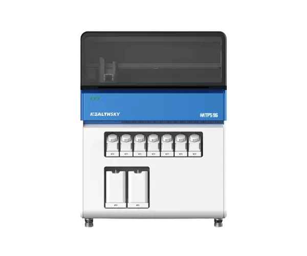
HealthSky's HATPS 96 Automated Tissue Processing System
 EN
EN
 jp
jp  ko
ko  fr
fr  de
de  pt
pt  ar
ar  es
es  ru
ru  fa
fa  id
id 


